

 schedule an appointment
Corneal Tissue Addition Keratoplasty (CTAK and CAIRS)
Keratoconus causes the cornea to thin and bulge forward into a cone shape, which blurs and distorts vision. Corneal tissue addition keratoplasty treats this by placing a precisely shaped piece of donor corneal tissue inside your cornea, where it flattens the cone and creates a smoother, more even curve. Unlike a corneal transplant, your own cornea stays in place, and none of it is removed.
There are two forms of tissue addition. CAIRS uses arc-shaped segments of donor tissue, and CTAK uses an inlay custom-designed from the maps of your cornea. Georgia Eye Partners was the first practice in Georgia to offer CAIRS, and we have more experience with corneal tissue addition than any other practice in the Atlanta area.
How CTAK and CAIRS Can Help
When the cornea has a more even shape, it can focus light more clearly. For most patients, tissue addition can lead to:
- Sharper vision in glasses or soft contact lenses
- A rigid or scleral lens that is easier to fit and more comfortable to wear
- Lower astigmatism, typically cut by about half in our patients
- Better best-corrected vision (the sharpest you see with glasses or contacts), typically one to three more lines on the eye chart in our patients
- Better vision with no correction at all: about four in five of our patients gain two or more lines on the eye chart
For some patients, tissue addition also makes a later procedure possible, such as topography-guided PRK, a toric implantable lens (ICL) or lens replacement, to further reduce the need for glasses or contacts.
A well-fitted specialty contact lens usually still gives the sharpest vision possible for a keratoconus cornea. Tissue addition is a good choice if you want to see well without a specialty lens, if you can’t wear one comfortably, or if you want the lens you wear to fit better.
How CTAK and CAIRS Work
Both procedures start the same way. A femtosecond laser, the same precise laser used in bladeless LASIK, creates a narrow channel inside the middle layer of the cornea, and the surgeon slides the donor tissue into that channel. Adding thickness in the right places flattens the steepest part of the cone and evens out the lopsided shape that blurs vision. How much the cornea flattens depends on the size, shape, position and depth of the tissue, and your surgeon plans each of these for your cornea.
CAIRS
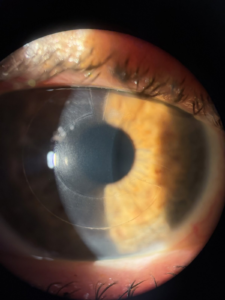
CAIRS stands for corneal allogeneic intrastromal ring segments: ring segments cut from donor cornea and placed in the cornea’s middle layer. The segments come from donor corneas supplied by an eye bank, either fresh or sterilized and shelf-stable. The surgeon places them in channels partway between the center and the edge of the cornea, choosing the length and thickness of each segment to suit your cornea.
 A CAIRS segment in place. It sits inside the cornea, outside the line of sight, and is hard to see without magnification.
CTAK
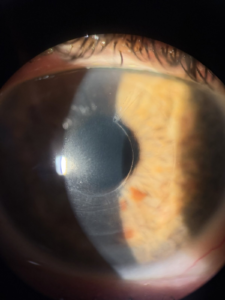
CTAK stands for corneal tissue addition keratoplasty. With CTAK, each inlay is designed for one eye. The surgeon sets its thickness, length and position from your corneal topography and tomography, and the eye bank cuts it to that design with a femtosecond laser. The tissue is sterilized and preserved, so it can be prepared ahead of surgery to your exact specifications. It is also clearer in the eye than a CAIRS segment, so it is nearly invisible once it has settled.
A CTAK inlay in place, lit from the side to make it visible.
Which Option is Right For You?
Both procedures work best when the center of the cornea is free of scarring. CAIRS is a great fit for most people, especially when the cone sits in the typical location. Because the segments come in set shapes, your surgeon chooses the length and thickness that best match your cornea. CTAK is often the better choice when the cone is in an unusual spot, has an uneven shape or is more advanced. Since each inlay is custom-designed for your eye, it can be tailored to these more complex cases.
After reviewing your corneal scans, your corneal surgeon will recommend the option that best fits your eye.
Why Donor Tissue Instead of Plastic Rings?
For about a decade, we treated keratoconus with INTACS, clear plastic ring segments that flatten the cornea in a similar way. However, plastic segments come in a limited range of sizes, and over time some can cause the tissue above them to thin and break down.
Donor tissue is natural corneal tissue, so it blends well with your own cornea. It can be cut to the size your eye needs, and CAIRS segments can be adjusted, exchanged or removed later if needed. In a head-to-head comparison, eyes treated with CAIRS were more likely to gain two or more lines of best-corrected vision than eyes treated with plastic segments.
If you have older INTACS that are causing problems, we can remove them and talk through your next steps.
Combining Tissue Addition With Crosslinking
Tissue addition reshapes the cornea, but it does not stop keratoconus from progressing.
If your keratoconus is getting worse, or hasn’t been shown to be stable, we pair tissue addition with corneal crosslinking. We sometimes add crosslinking in a stable eye too, because it can add to the flattening.
We place the tissue first and crosslink two to four weeks later. Crosslinking stiffens the cornea, and a cornea that has already been stiffened may not reshape as much when the tissue goes in. The crosslinking is done with Epioxa, which leaves the cornea’s surface layer in place.
Who is a Candidate for CTAK and CAIRS?
You may be a good candidate for tissue addition if:
- You have keratoconus, pellucid marginal degeneration (a thinning near the lower edge of the cornea), or ectasia (a similar bulging after LASIK, PRK or RK)
- The center of your cornea is clear
- Glasses or soft contacts no longer give you acceptable vision, or specialty lenses are hard to fit or uncomfortable
- Your cornea is thick enough where the tissue will sit
Patients with mild to moderate keratoconus tend to see the best results, especially when their vision with glasses is still fairly good but no longer good enough for daily life.
Tissue addition is also an option for many people who aren’t candidates for laser treatments like topography-guided PRK. During your evaluation, we’ll review your scans and let you know whether CTAK or CAIRS is a good fit for your eyes.
What to Expect From CTAK and CAIRS
Your first visit includes a full keratoconus evaluation with corneal tomography. If CTAK is the plan, we use those scans to design your inlay, and surgery is scheduled once the tissue has been prepared.
Tissue addition is an outpatient procedure done with numbing drops. You’ll feel some pressure, with little or no pain. Afterward, the eye may feel scratchy or watery for a day or two, and you’ll use eye drops for a few weeks.
Many patients notice some improvement as soon as the day after surgery. Most are back in contact lenses about two weeks later. Vision keeps changing over the following weeks to months as the cornea settles into its new shape. Once it is stable, we update your glasses or refit your contact lenses.
FAQs About Corneal Tissue Addition Keratoplasty
Schedule a Consultation
If keratoconus is making it hard to see clearly in glasses or wear contact lenses comfortably, tissue addition may help restore a smoother, more even cornea and sharper vision. At your consultation, we’ll take detailed scans of your cornea, talk through your goals and let you know whether CTAK or CAIRS is a good fit for your eyes.
Request an appointment online or call (404) 531-9988. Bring any earlier corneal scans, your glasses, and your contact lenses if you wear them.
References
- Jacob S, et al. Corneal allogenic intrastromal ring segments (CAIRS) combined with corneal cross-linking for keratoconus. J Refract Surg. 2018;34(5):296-303. doi:10.3928/1081597X-20180223-01
- Greenstein SA, et al. Corneal tissue addition keratoplasty: new intrastromal inlay procedure for keratoconus using femtosecond laser-shaped preserved corneal tissue. J Cataract Refract Surg. 2023;49(7):740-746. doi:10.1097/j.jcrs.0000000000001187
- Kozhaya K, et al. Management of anterior stromal necrosis after polymethylmethacrylate ICRS. J Refract Surg. 2022;38(4):256-263. doi:10.3928/1081597X-20220223-01
- Asfar KE, et al. CAIRS versus synthetic segments: a single segment comparative analysis using propensity score matching. J Refract Surg. 2024;40(11):e863-e876. doi:10.3928/1081597X-20241002-02
- Keskin Perk FFN, et al. Long-term results of sterile corneal allograft ring segments implantation in keratoconus treatment. Cornea. 2025;44(4):475-482. doi:10.1097/ICO.0000000000003592


